
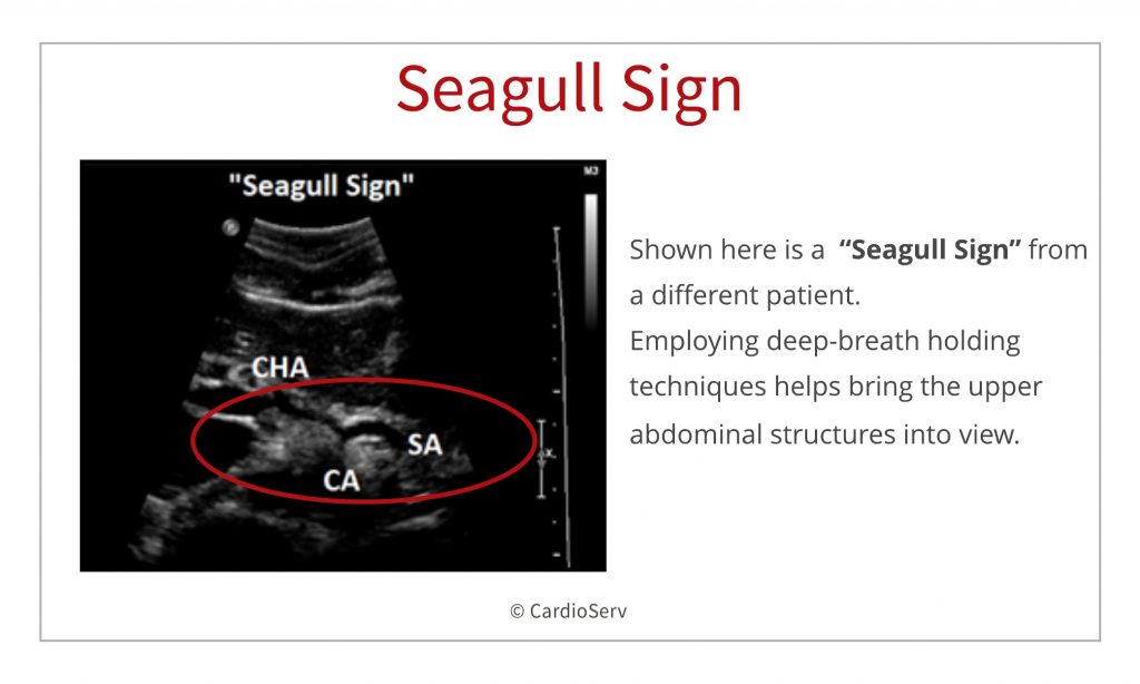
The infamous “Seagull Sign” 🦅 😍 The “seagull sign” is referred to the celiac trunk and its
Ultrasound is a useful modality to quickly assess the aorta for the presence of an aneurysm, or arterial dissection, and to determine the relative risk of impending rupture. To do this, it is important for an ultrasonographer to learn how to accurately image and measure the diameter of an aortic aneurysm.

The infamous “Seagull Sign” 🦅 😍The “seagull sign” is referred to the celiac trunk and its
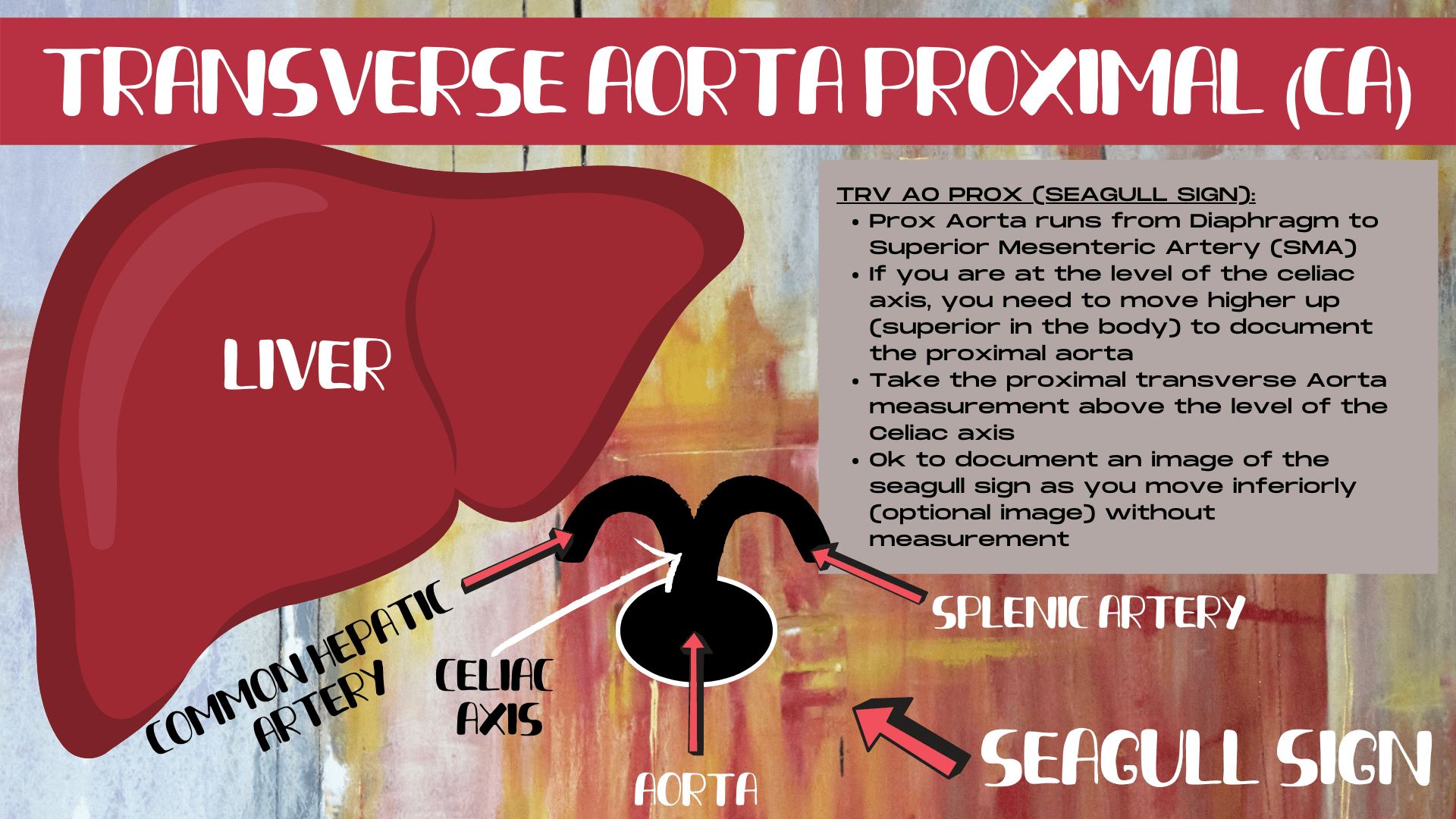
Celiac trunk with seagull sign: Arising from the aorta (A) is the celiac trunk (C), branching into the hepatic artery (B) and splenic artery (D) Figure 7.3 Superior mesenteric artery: Superior mesenteric artery (A) is the second main branch from the abdominal aorta, just inferior to the celiac trunk Figure 7.4
Obstetric Ultrasound Signs YouTube
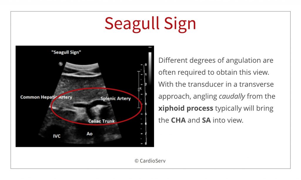
Look for the "seagull sign" - the wings are the hepatic (screen left) and splenic (screen right) arteries. The left gastric artery, the third component of the celiac trunk is usually not visualized on ultrasound.
Abdominal Ultrasound for Echocardiographers Branches of the Aorta Cardioserv
The gull-wing appearance , also known as seagull erosions or sawtooth appearance , is classically seen in erosive osteoarthritis, typically on posteroanterior radiographs of the hands, although has also been reported in psoriatic and rheumatoid arthritis.
AORTA ULTRASOUND STOP DOING THESE 5 THINGS Sonography Minutes
Transverse abdominal ultrasound image demonstrating the bifurcation of the celiac artery (CA) into the hepatic artery (HA) on the right and splenic artery (SA) on the left, resembling a seagull. The asterisk (*) denotes the abdominal aorta Full size image Fig. 2 A seagull stretching its wings [ 2] Full size image

Ultrasound Seagull Sign (English) YouTube
Seagull Sign Take-off of the celiac trunk and bifurcation to hepatic and splenic arteries Bladder ultrasound Twinkle Sign Rapid alternation of color immediately behind a stationary echogenic object, acquiring a false appearance of movement Can indicate stone in the UVJ Cardiac ultrasound McConnell's Sign

Biliary Images Emergency Ultrasonography
. 3 This has also been described as a "whale's tail"sign. 4 The seagull appearance has also been described as a sign to recognise the pulmonary arteries in the fetus. 5 Other vascular.

Seagull sign or Gull wing appearence Pet ct, Gull, Radiology
The most commonly used definition of an AAA is a maximum infrarenal abdominal aortic diameter greater than 3 cm on ultrasound or CT. [ 8, 9, 10] POCUS performed by a physician at the bedside is.
Abdominal Ultrasound for Echocardiographers Branches of the Aorta Cardioserv
The arterial bifurcation most commonly described as having a seagull appearance is the transverse ultrasound appearance of the coeliac bifurcation into the common hepatic artery and splenic artery.1 This appearance was recognised from the early days of ultrasound both in the adult by 1979,2 and in the fetus by 1984.3 This has also been described.

Trunchiul celiac . "Semnul pescarusului"/ "Seagull sign" YouTube
The "seagull sign" in abdominal ultrasound identifies the celiac trunk and its division into hepatic and splenic arteries creates the wings of the seagull.[6] Wherever an artery divides.

Seagulls of endoscopic ultrasound. Abstract Europe PMC
The "seagull sign" in abdominal ultrasound identifies the celiac trunk and its division into hepatic and splenic arteries creates the wings of the seagull. Wherever an artery divides into two smaller arteries or two veins unite to form a larger vein, they can give an appearance of a seagull. The union of the splenic vein, superior.

3 IVC Ultrasound Prereading for FCUS Course Intensive Care Network
Here's the schematic of that transverse view of that first branch, the celiac artery, sometimes call this the seagull sign, it looks like the wings of a seagull. You see the common hepatic artery to the right and the splenic artery to the left. There is also the left gastric branch of the celiac which you often don't see that well on.

Ultrasonographic images of eyes of representative dogs with intraocular... Download Scientific
This week we will help you easily identify with ultrasound the branches of the aorta and other vasculature, SMA, Celiac Axis, Renal Veins and Arteries.. These 2 branches often create what is known as the "Seagull Sign". This sign can be very useful in identifying the visceral vessels. Step 2: Identify other visceral vessels

AAA — MMHEME
The major branches of the aorta often have characteristic sonographic appearances. The axial view of the celiac axis originating from the aorta is commonly known as a seagull sign ( Fig. 20.4, Video 20.2). The axial view of the SMA takeoff surrounded with hyperechoic fatty tissue is known as the Mantel clock sign.

A 61yearold man presented with acetabular fracture of the right... Download Scientific Diagram
Seagull appearance: Vascular anatomy on ultrasound. J Med Imaging Radiat Oncol. 2018 Oct;62 Suppl 1:104. doi: 10.1111/1754-9485.48_12784.

Signe de la mouette EchoUrgences
0:00 / 3:43 • Ultrasound Seagull Sign (English) Shakil Health Society 2.02K subscribers Subscribe 26 Share Save 741 views 2 years ago #celiac #ultrasound #vascularultrasound Ultrasound Seagull.